Early & Late Hours

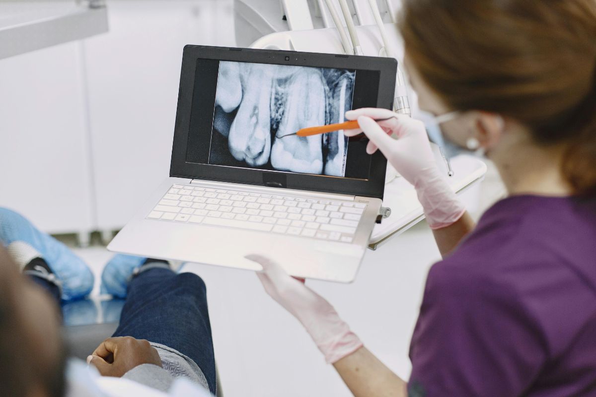
At first glance, a healthy-looking smile can seem like proof that everything is fine. But a digital x-ray vs visual exam comparison tells a very different story, because plenty of dental problems start out completely invisible to the naked eye. At Evergreen Dental Group, tucked into the community of Evergreen, Colorado, our team relies on both tools together, not because one is outdated, but because each one sees something the other can’t.
So what exactly does a visual exam miss, and why does that gap matter for your long-term oral health? Let’s break it down.
What a Visual Exam Can Actually Tell Your Dentist
A visual exam is still an essential first step. With a mirror, good lighting, and trained eyes, a dentist can spot a lot:
- Visible cavities on the surface of a tooth
- Cracked, chipped, or worn enamel
- Signs of gum inflammation or recession
- Discoloration or staining
- Obvious plaque and tartar buildup
This kind of exam is fast, painless, and genuinely useful, and it’s a core part of what your trusted dentist checks at every visit. The problem is that it can only assess what’s on the surface, which leaves a lot of territory unexamined.
The Blind Spots of a Visual-Only Check
Teeth are three-dimensional, and a huge portion of what matters is hidden from view. Consider what a visual exam simply cannot reach:
Between the teeth. Cavities that form in the tight contact points between two teeth are nearly impossible to catch just by looking, since the surfaces are pressed together.
Beneath old fillings. Decay can develop underneath an existing filling or crown without changing how the restoration looks from the outside.
Below the gumline. Bone loss, infection, and early-stage gum disease often start beneath the tissue, where eyes alone can’t follow.
Inside the tooth. Cracks that run below the enamel, or infections forming near the roots, rarely show any visible surface sign until they’ve become painful.
Where Digital X-Rays Fill the Gap
This is exactly the territory digital x-rays are built for. Instead of relying on what’s visible, X-rays capture images of the internal structure of your teeth and the bone supporting them, revealing:
- Decay forming between teeth, long before it reaches the surface
- Infections or abscesses developing near the tooth root
- Bone density and levels, which matter for gum health and implant planning
- The position of unerupted or impacted teeth, including wisdom teeth
- Cracks or fractures that aren’t visible externally
Because digital X-rays use significantly less radiation than older film-based X-rays and produce images almost instantly, they’ve become a routine, low-impact part of a thorough exam rather than a rare or invasive step.
Why Using Both Together Makes the Difference
Neither tool replaces the other. A visual exam gives immediate, real-world context, while X-rays fill in what’s happening beneath the surface. Used together, they give your dental team a complete picture instead of half of one.
This combination matters most for catching problems early. A cavity found while it’s still small and confined to enamel is a simple fix. The same cavity, left undetected for a year or two because it was hiding between teeth, can grow into something that needs a root canal or extraction.
How Often Should You Get Digital X-Rays?
There’s no single answer that fits everyone, since it depends on your age, oral health history, and risk factors. That said, general guidelines many dental teams follow include:
- Bitewing X-rays roughly once a year for patients at average risk of cavities
- Less frequent imaging for patients with excellent oral health and low risk
- More frequent imaging for patients with a history of decay, gum disease, or ongoing dental work
Your dentist will typically tailor this schedule to your specific situation rather than applying a one-size-fits-all rule.
Addressing the “Is It Safe?” Question
It’s a fair concern, and one patients bring up often. Modern digital X-rays use a very small amount of radiation, comparable to what you’d naturally encounter during a short flight or a single day of background exposure. Lead aprons and thyroid collars add an extra layer of protection during the process, making digital imaging a safe, well-established part of preventive dental care.
Final Thoughts
A confident smile in the mirror doesn’t always mean everything underneath is healthy. Between-teeth decay, bone changes, and hidden infections rarely announce themselves visually until they’ve progressed. Pairing a visual exam with digital X-rays gives your dental team the fuller picture needed to catch issues while they’re still small, simple, and inexpensive to treat.
Curious what a full picture of your oral health looks like? Book a visit at our Evergreen, CO office, where our team combines a careful visual exam with digital X-ray imaging to catch what matters most, before it becomes a bigger problem. Request an appointment with Evergreen Dental Group today.



New Patients and Emergency Appointments are Welcome!